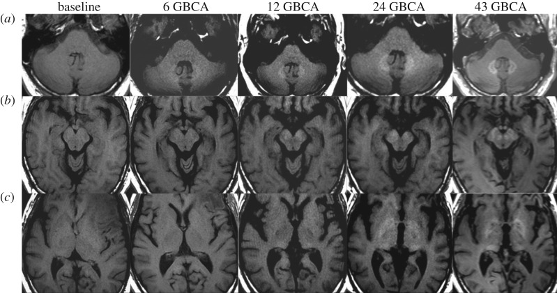
Figure 2.
MR images in a 52-year-old man with left frontal glioblastoma. Dentate nucleus (a), cerebral peduncle, substantia nigra and red nucleus (b), and globus pallidus and posterior thalamus (c) all gradually increase unenhanced T1 signal intensity after 43 linear GBCA administrations. Adapted from [14].

