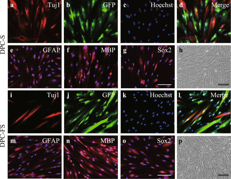
Figure 1.
Morphology and expression of neural marker proteins of DPCs. DPCs were transfected with GFP reporter gene using a lenti-viral vector and cultured in the absence or presence of FGF2 (a–h. DPC-S or i–p. DPC-FS, respectively). Cultured DPCs were characterized immunohistochemically using antibodies against GFP (green; b,d,j,l) and/or each indicated neural cell marker (red), respectively: anti-Tuj1- (a,d,i,l), anti-GFAP- (e,m), and anti-MBP- (f,n) and anti-Sox2- (g,o) antibody, respectively. Nuclei were stained with Hoechst 33342 (blue; c–g,k–o). All DPCs showed spindle-shaped fibroblast morphologies (h,p). Scale bar, 100 μm; double bars, 200 μm.

