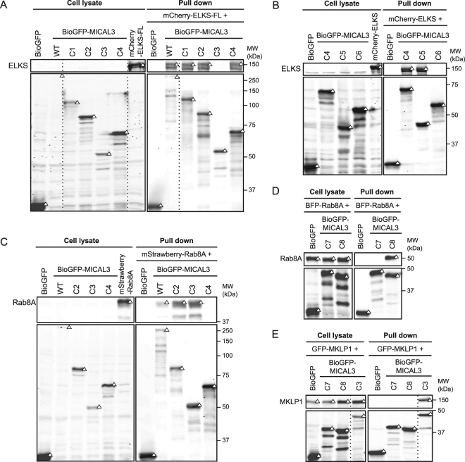
Figure 3.
Characterization of the domains responsible for the interactions between MICAL3, ELKS and Rab8A. Results of streptavidin pull down assays. In all panels, triangles indicate signals at the expected molecular weight. Dashed lines indicate cropped lanes irrelevant for this study. Full images are presented in Supplementary Figure S3. (A,B) Pull downs are performed by mixing the lysates of cells co-expressing biotin ligase BirA and differently truncated BioGFP-tagged MICAL3, with the lysates of cells expressing mCherry-ELKS-FL. Rabbit-anti-ELKS antibody and mouse-anti-GFP antibody were used for western blotting, depicted in the upper and lower boxes respectively. (C) Illustrated pull down is performed by mixing the lysates of cells co-expressing biotin ligase BirA and differently truncated BioGFP-tagged MICAL3, with the lysates of cells expressing mStrawberry-Rab8A. The blot is incubated with rabbit-anti-Rab8A and mouse-anti-GFP antibodies for the upper and lower boxes. (D) The pull down is performed by mixing the lysates of cells co-expressing biotin ligase BirA and indicated BioGFP-tagged MICAL3 constructs with lysates of cells expressing BFP-Rab8A. Western blot is performed using rabbit-anti-Rab8A and mouse-anti-GFP antibodies for the upper and lower boxes. (E) Pull down is performed by the mixing of lysates of cells co-expressing biotin ligase BirA with indicated BioGFP-tagged proteins with lysates of cells expressing GFP-MKLP1. Mouse-anti-GFP antibody is used in both upper and lower boxes.

