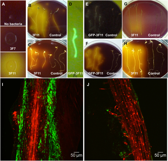
Figure 2.
Testing endophyte 3F11 for its ability to cause ambient acidification using bromocresol purple as a pH indicator. A change in color from purple to yellow indicates a decline in pH. (A) Test for in vitro acid production by a representative colony of 3F11 using NBRIP agar after 24 h compared to a negative control endophyte, 3F7. (B,C) Test for in planta acid production of 3F11 following seed inoculation of annual ryegrass. Shown are three 3F11-inoculated roots (left) or uninoculated roots (right) placed on insoluble-P agar medium (NBRIP) supplemented with bromocresol purple and either (B) no kanamycin or (C) kanamycin. The growth of 3F11 but not annual ryegrass was previously shown to be suppressed by kanamycin. (D) Confirming the colonization of GFP-tagged 3F11 cells on annual ryegrass roots on an LB agar plate following coating onto seeds. (E,F) Testing the co-localization of 3F11 cells with acid production in planta. Shown are three GFP-3F11-inoculated roots (left) or uninoculated roots (right) placed on insoluble-P agar medium (NBRIP) supplemented with bromocresol purple and kanamycin and (E) imaged using UV light to reveal GFP or (F) white light to reveal acid production (yellow). (G,H) Test for the effect of P-bioavailability in the rhizosphere on the in planta acid production of 3F11. Shown are three GFP-3F11-inoculated roots (left) or uninoculated roots (right) placed on (G) soluble-P agar medium supplemented with bromocresol purple and (H) insoluble P containing agar medium (NBRIP) supplemented with bromocresol purple. (I,J) Representative confocal microscopy images showing colonization of GFP-tagged 3F11 cells on annual ryegrass root systems 7 days following seed-inoculation and growth on either (I) rock P, or (J) soluble P, showing the difference in the extent of colonization.

