Figure 2.
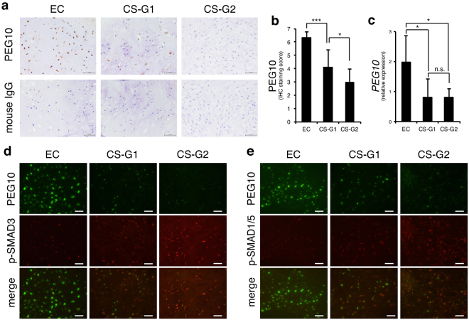
PEG10 accumulates in enchondromas but is eliminated in chondrosarcomas. (a) IHC of PEG10 in enchondroma and chondrosarcoma specimens. Normal mouse IgG was used as a negative control. EC, enchondromas (n = 7); CS-G1, grade 1 chondrosarcomas (n = 11); CS-G2, grade 2 chondrosarcomas (n = 7). Scale bar = 50 μm. (b) Summed score of the percentage and intensity of positive staining in IHC. (c) Expression of PEG10 was examined by RT-qPCR. EC (n = 6); CS-G1 (n = 5); CS-G2 (n = 6). *P < 0.05; ***P < 0.001; n.s., not significant. (d,e) Double immunofluorescence (IF) of PEG10 and p-SMAD3 or p-SMAD1/5 in enchondroma and chondrosarcoma specimens. Signals of PEG10 were detected by an anti-mouse Alexa Fluor 488 antibody (green), while p-SMADs were stained by an anti-rabbit Alexa Fluor 568 antibody (red). Scale bar = 25 μm.

