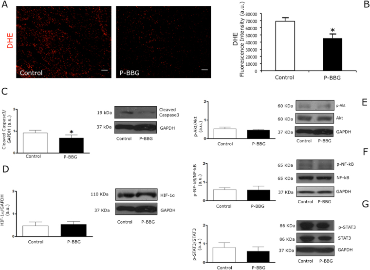
Figure 5.
Effects of P-BBG diet on levels of myocardial oxidative stress, cleaved caspase-3, HIF-1α, pAkt/Akt, pSTAT3/STAT3 and pNF-kB/NF-kB. (A) Representative images of DHE (dihydroethidium) staining of Control and P-BBG left ventricles. (B) P-BBG diet (n = 15) prevented an increase in anion superoxide generation following cardiac ischemia/reperfusion injury compared to Control diet (n = 15); the results are shown as arbitrary units of fluorescence intensity (see Methods). (C) The myocardial levels of cleaved caspase-3 were lower in P-BBG (n = 7) than Control (n = 7) hearts. Levels of cleaved caspase-3 are expressed as arbitrary units of cleaved caspase-3 (19 kDa, MW)/GAPDH (37 kDa, MW) ratio. Representative images of cropped densitometric bands of cleaved caspase 3 AND GAPDH are shown. The full-length blots/gels are presented in Supplementary Figure 1C. (D) The myocardial levels of HIF-1α are similar in P-BBG (n = 7) and Control (n = 7) hearts. Levels of HIF-1α are expressed as arbitrary units of HIF-1α (110 kDa, MW)/GAPDH (37 kDa, MW) ratio. Representative images of cropped densitometric bands of HIF-1α and GAPDH are shown. The full-length blots/gels are presented in Supplementary Figure 1C. (E) The myocardial levels of p-Akt/Akt ratio are similar in P-BBG (n = 7) and Control (n = 7) hearts. Levels of p-Akt (60 kDa, MW)/Akt (60 kDa, MW), normalized to GAPDH levels, are expressed as arbitrary units. Representative images of cropped densitometric bands of p-Akt, Akt and GAPDH are shown. The full-length blots/gels are shown in Supplementary Figure 1C. (F) The myocardial levels of p-NF-kB/NF-kB ratio are similar in P-BBG (n = 7) and Control (n = 7) hearts. Levels of p-NF-kB (65 kDa, MW)/NF-kB (65 kDa, MW), normalized on GAPDH levels, are expressed as arbitrary units. Representative images of cropped densitometric bands of p-NF-kB, NF-kB and GAPDH are shown. The full-length blots/gels are presented in Supplementary Figure 1D. (G) The myocardial levels of p-STAT3/STAT3 ratio are similar in P-BBG (n = 7) and Control (n = 7) hearts. Levels of p-STAT3 (86 kDa, MW)/STAT3 (86 kDa, MW), normalized on GAPDH levels, are expressed as arbitrary units. Representative images of cropped densitometric bands of p-STAT3, STAT3 and GAPDH are shown. The full-length blots/gels are presented in Supplementary Figure 1E. P-BBG: low fat diet supplemented with barley beta-D-glucan enriched pasta (3 g/100 g of dry weight); HIF-1α: hypoxia-inducible factor 1-alpha; GAPDH: glyceraldehyde 3-phosphate dehydrogenase; p-Akt: phospho-Akt; p-NF-kB: phospho- nuclear factor Kappa b; p-STAT3: phospho- signal transducer and activator of transcription 3. All measurements are mean ± SD. *p < 0.05 vs. Control.

