Figure 7.
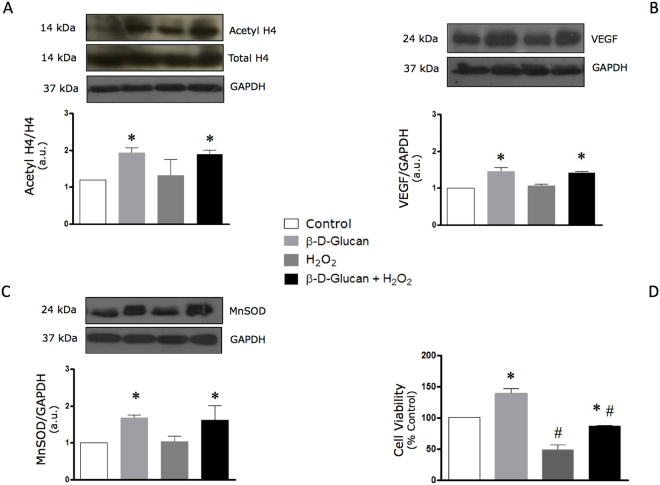
Acute oxidative stress does not affect levels of H4 histone acetylation, VEGF and MnSOD in viable endothelial cells chronically treated with barley β-D-glucan. (A) 7-day treatment with 3% barley β-D-glucan (BBG) increases the endothelial levels of acetyl H4 histone compared to untreated HUVECs, which are not affected by acute oxidative stress. Levels of acetyl H4 (14 kDa, MW)/H4 (14 kDa, MW), normalized on GAPDH levels, are expressed as arbitrary units. Representative images of cropped densitometric bands of acetyl H4, H4 and GAPDH are shown. The full-length blots/gels are presented in Supplementary Figure 2A. (B) The protein levels of VEGF in HUVECs increase after long-term exposure to 3% BBG compared to untreated cells at rest and during acute oxidative stress. Levels of VEGF are expressed as arbitrary units of VEGF (24 kDa, MW)/GAPDH (37 kDa, MW) ratio. Representative images of cropped densitometric bands of VEGF and GAPDH are shown. The full-length blots/gels are presented in Supplementary Figure 2B. (C) The protein levels of MnSOD in HUVECs are increased after long-term exposure to 3% BBG compared to untreated cells, yet are not altered after 1 h exposure to hydrogen peroxide (see Methods). Levels of MnSOD are expressed as arbitrary units of MnSOD (24 kDa, MW)/GAPDH (37 kDa, MW) ratio. Representative images of cropped densitometric bands of MnSOD and GAPDH are shown. The full-length blots/gels are presented in Supplementary Figure 2C. (D) BBG promotes the endothelial cell survival after 1 hour treatment with 400 μM H2O2. HUVECs: human umbelical vascular endothelial cells; H4: histone H4; VEGF: vascular endothelial growth factor; MnSOD: manganese superoxide dismutase; GAPDH: glyceraldehyde 3-phosphate dehydrogenase; H2O2: hydrogen peroxide. All measurements are mean ± SD. *p < 0.05 vs. untreated condition; #p < 0.05 vs. resting condition.

