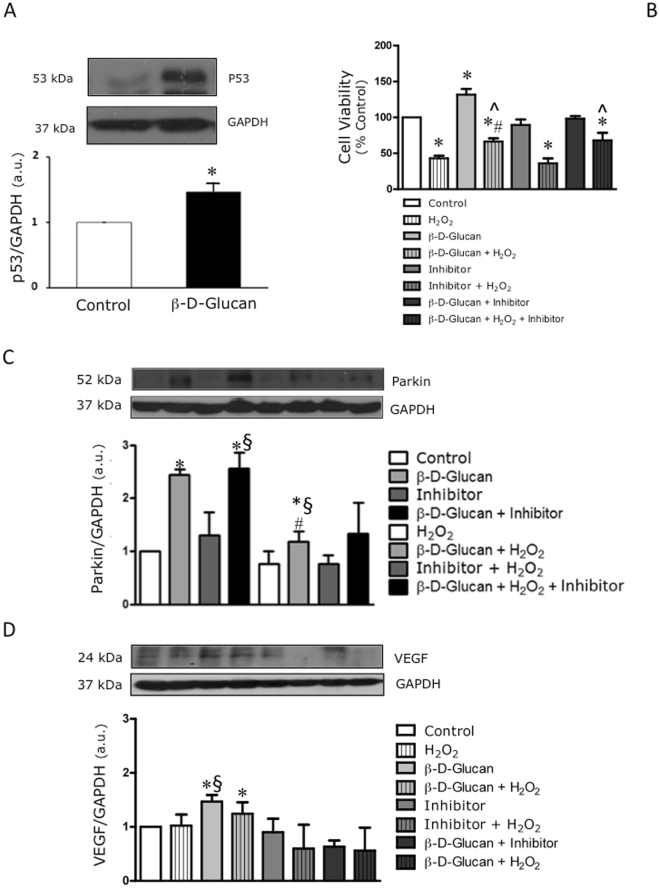
Figure 8.
BBG induces the protein expression of Parkin independently of p53 activity. (A) 7-day treatment with 3% barley β-D-glucan (BBG) increases the endothelial levels of p53 compared to untreated HUVECs. Levels of p53 are expressed as arbitrary units of p53 (53 kDa, MW)/GAPDH (37 kDa, MW) ratio. Representative images of cropped densitometric bands of p53 and GAPDH are shown. The full-length blots/gels are presented in Supplementary Figure 2D. (B) The sustained co-treatment of HUVECs with 3% BBG and PFT-α (Inhibitor) does not impair cell viability. BBG preserves the viability of H2O2-stressed cells, but not Inhibitor. (C) The sustained treatment of HUVECs with 3% BBG increases the protein levels of Parkin compared to untreated cells (Control). The co-treatment with PFT-α does not affect the BBG-induced protein expression of Parkin. In addition, the decay of Parkin protein expression in stressed BBG-treated cells is smaller than in stressed untreated cells. (D) HUVECs co-treated with 3% BBG and PFT-α show lower protein levels of VEGF. GAPDH: glyceraldehyde 3-phosphate dehydrogenase; PFT-α: pifithrin-alpha; H2O2: hydrogen peroxide; VEGF: vascular endothelial growth factor. All measurements are mean ± SD. *p < 0.05 vs. untreated condition; §p < 0.05 vs. Inhibitor; #p < 0.05 vs. corresponding resting condition; ^p < 0.05 vs H2O2.

