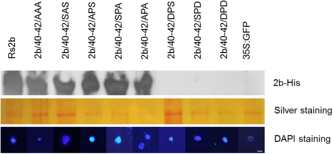
Figure 6.

Western blot analysis of the Rs2b and the different mutants from cell nuclei purified from infiltrated N. benthamiana leaves. His-tagged 2b proteins were detected with penta-his antibody and silver staining was used to monitor the equivalence of protein loading. Mutants SPS/40-42/AAA, SPS/40-42/APA, and single mutants SPS/40-42/APS, SPS/40-42/SPA and SPS/40-42/SAS were detectable from purified nucleoli with penta-his antibody. The mutants 2b/40-42/DPS, 2b/40-42/SPD and 2b/40-42/DPD were not detectable in nuclei. DAPI-staining was used for visualisation of the purified nuclei (appearing blue) with fluorescence microscope using UV-light. The bar represents 10 µm.
