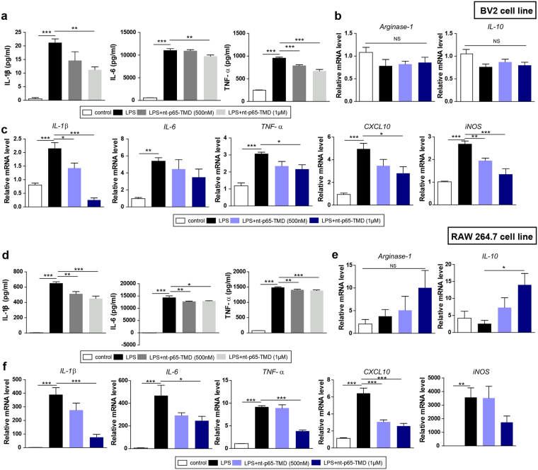
Figure 6.
Treatment with nt-p65-TMD reduces inflammatory mediators in BV2 microglia and RAW264.7 macrophages and elevates anti-inflammatory mediators in the RAW264.7 macrophages. (a) The culture supernatants from the BV2 cell line were collected 24 h after LPS stimulation and the IL-1β (7 repeats), IL-6 (7 repeats), and TNF-α (7 repeats) levels were evaluated by ELISA. (b) The transcripts of anti-inflammatory mediator genes such as arginase-1 (7 repeats) and IL-10 (7 repeats) and (c) pro-inflammatory mediator genes such as IL-1β (4–5 repeats), IL-6 (6 repeats), TNF-α (6–7 repeats), CXCL10 (7 repeats), and iNOS (7 repeats), of BV2 microglia were measured by real-time PCR. (d) The culture supernatants from the RAW264.7 cell line were collected 24 h after LPS stimulation, and the IL-1β (7 repeats), IL-6 (5 repeats), and TNF-α (5 repeats) levels were assessed by ELISA. (e) The transcripts of anti-inflammatory mediator genes, such as arginase-1 (5–6 repeats) and IL-10 (5 repeats), and (f) pro-inflammatory mediator genes, such as IL-1β (5–7 repeats), IL-6 (5 repeats), TNF-α (7 repeats), CXCL10 (7 repeats), and iNOS (6 repeats), of RAW 264.7 macrophages were calculated by real-time PCR. Values are means ± SEM. P values were calculated with multiple comparisons by Bonferroni tests. *p < 0.05, **p < 0.01, ***p < 0.001 vs. LPS. Statistical parameter (Supplementary Table 7).

