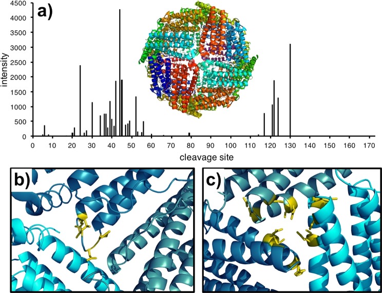
Figure 4.
Yields of c-type fragment ions plotted with respect to their cleavage site (a) with the X-ray crystal structure of ferritin (PDB: 1IER) comprised of all L-subunits (shown in different colors). Mapping the major fragment ions from NECD onto the crystal structure for sites 43–53 (b) indicates that the cleavages are centered in the helix-loop region. Fragment ions from sites 114–130 (c) instead indicate cleavages from the 3-fold axis pore.

