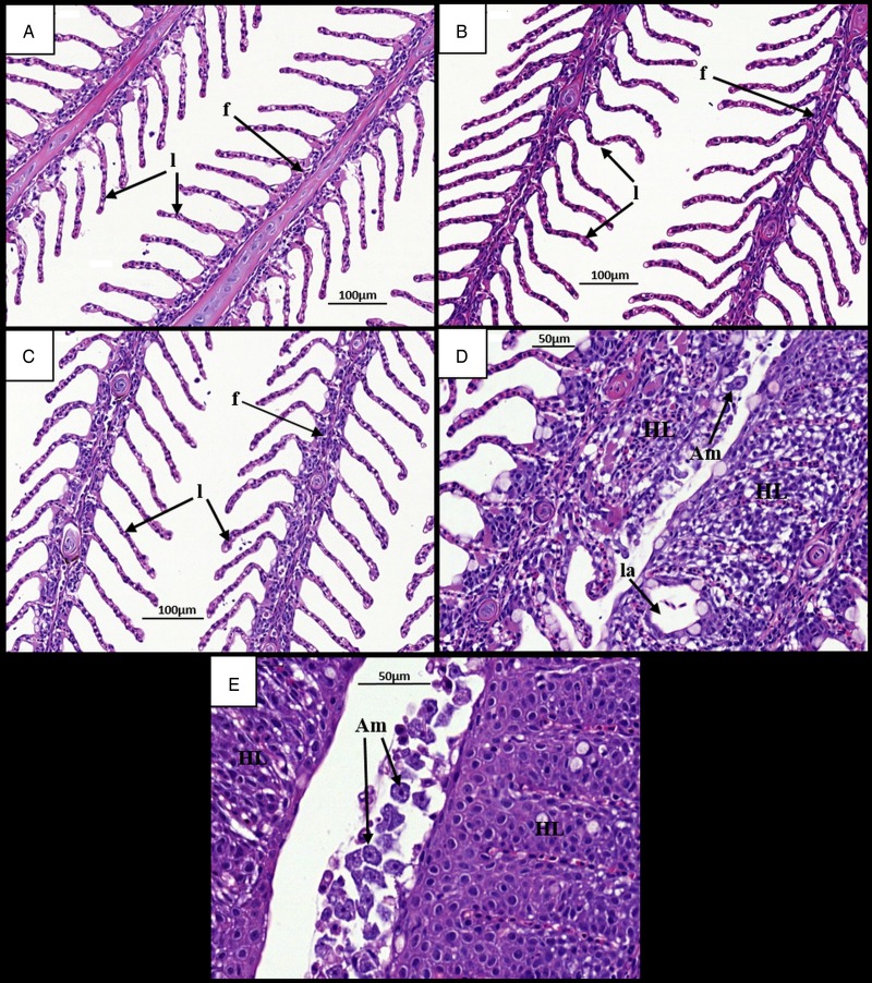
Fig. 2.
Time changes in histological pathology in the gills of infected and uninfected diploid Atlantic salmon. (A) Uninfected fish at 7 dpi. Individual filaments (f) and lamellae (l) visible. (B) Infected fish at 7 dpi. (C) Uninfected fish at 28 dpi. Individual filaments and lamellae remain visible. (D) Infected fish at 28 dpi. Hyperplastic AGD lesions (HL) clearly visible with lamellar fusion causing lacunae formation (la). Amoeba (Am) visible next to lesion. (E) Infected fish at 28 dpi. Numerous amoebae associated with hyperplastic gill tissue. AGD, amoebic gill disease.

