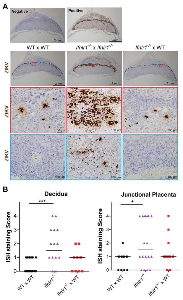
Figure 4. In situ hybridization of placentas from ZIKV-infected Ifnlr1−/− and pegIFN-λ2-treated mice.
A. Representative ISH staining. Top panels. ISH staining with negative (bacterial gene dapB) and positive (mouse Ppib gene) probe controls. Middle panels. Images showing ZIKV RNA localization in placental and decidual tissues from dams after matings (dam and sire genoypes are listed in order, respectively). Low and high power images are presented in vertical sequence. In low power images, solid black lines outline the boundary between maternal decidua and fetal placenta. Images of decidua and placenta within the red and blue frames (top boxes) are shown at higher magnification (two lower boxes). The images are representative of several sections from independent experiments. B. Grading of ISH staining intensity. ISH staining intensities were assessed in a blinded manner on a scale of 0 to 4, and data analyzed by Kruskal-Wallis with Dunn’s post-test: *, P < 0.05; ***, P < 0.001. See also Figure S2.

