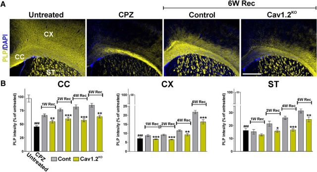
Figure 5.
Reduced PLP expression in the remyelination of the Cav1.2KO brain. A, Representative coronal sections of brain tissue immunostained for PLP collected from untreated animals (untreated), mice treated with CPZ for 7 weeks (CPZ), and control and Cav1.2KO mice at 6 weeks of recovery (6W Rec). Scale bar, 180 μm. B, Integrated fluorescence intensity for PLP was quantified in the lateral corpus callosum (CC), cingulate cortex (CX), and striatum (ST). Comparisons between experimental groups were made by the unpaired t test. Data represent pooled results from at least six brains per experimental group and values are expressed as mean ± SEM. ###p < 0.001 versus untreated; *p < 0.05, **p < 0.01, ***p < 0.001 versus control.

