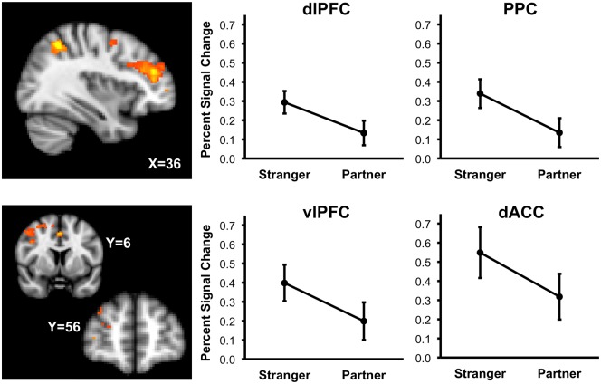
Fig. 2.
Probability maps depicting areas where percent signal change in the threat minus safe contrast is greater during Stranger handholding than Partner handholding. Sagittal slice (X = 36) depicts areas of dorsal lateral prefrontal cortex (dlPFC) and posterior parietal cortex (PPC). Coronal slices (Y = 6) depict areas of the anterior cingulate cortex (ACC) and the middle frontal gyrus. Graphs depict average threat minus safe percent signal change across stranger and partner handholding conditions, with 95% confidence intervals, in the dlPFC, PPC, vlPFC and dACC.

