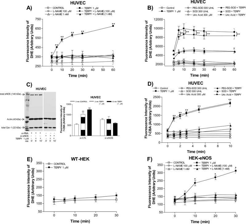
Figure 3. TERPY-induced eNOS dysfunction in HUVECs and HEK-eNOS.

Reactive oxygen species were measured using dihydroethidium (DHE) in human umbilical vein endothelial cells (HUVECs) (A,B), wild-type human embryonic kidney 293 cells (HEK-WT) (E) and HEK cells stably transduced with eNOS cDNA (HEK-eNOS) (F) in absence (Control) or presence of TERPY (1 μM) for times indicated. (C) and (D) Peroxynitrite was measured using coumarin-7-boronic acid (7-CBA) in HUVECs. (C) Representative Western blot of total eNOS, actin, and total Cav-1 in control si-RNA (right lane) and eNOS si-RNA treated cells (left lane). (A) and (F) L-NAME (100 μM or 1 mM) inhibited the TERPY-stimulated fluorescence intensity of DHE. (B) and (D) SOD (300 U/mL) and PEG-SOD (300 U/mL) decreased the fluorescence intensity of DHE and 7-CBA stimulated by TERPY, but not uric acid (300 μM). *p<0.05 indicates difference between TERPY, TERPY + SOD or TERPY + PEG-SOD versus basal condition (control, n=3–5) and **p<0.01 indicates difference between TERPY or TERPY + uric acid versus basal condition (control, n=3–5). “n” indicates independent experiments.
