. 2017 Sep 26;18(3):149–154. doi: 10.7181/acfs.2017.18.3.149

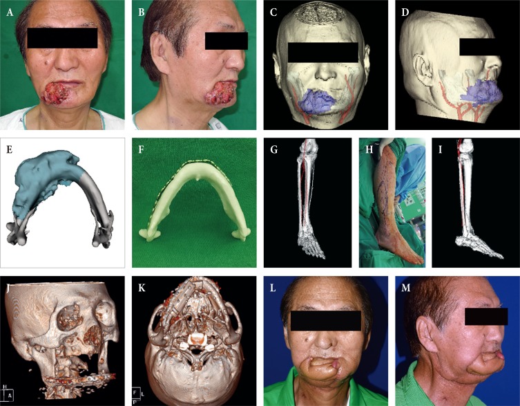
Fig. 1. (A, B) Computer-aided reconstruction of an extensive composite mandibular defect. A 67-year-old male patient with invasive squamous cell carcinoma on the right lower lip. (C, D) A transparent view of the tumor invading the mandibular bone with the vascular pedicle candidates for reconstruction. (E) The tumor-invading mandible was reconstructed as a three-dimensional (3D) object and the resection extent was estimated (dark cyan). (F) Given this estimation, an actual scale rapidly-prototyped patient mandible was manufactured for prefabrication of the reconstruction plate in order to shorten the operation time. (G) For precise preoperative planning and measurement of length, the image slicing plane was calibrated to a plane containing the axis of the donor fibular bone and peroneal artery, and the images were reconstructed as a 3D object. (H) Intraoperative markings and (I) the surgeon's view of the pedicle while harvesting the fibular flap. (J, K) A postoperative 3D view taken four weeks after mandibular reconstruction showing excellent restoration of the mandibular contour and continuity. (L, M) Six-month follow-up view of the patient after adjuvant radiation therapy.

Fig. 1