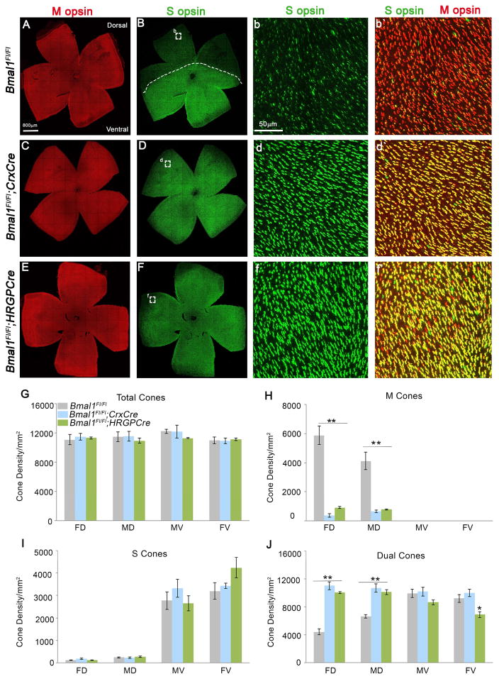
Figure 1. Bmal1 is required for maintenance of S opsin gradient.
(A–F) P24 retinal flat mount preparations coimmunolabeled with S opsin (green) and M opsin (red) from control (A and B) and Bmal1 conditional mutants (C–F). (B) Dotted line separating low S opsin expressing dorsal (superior) region from S opsin enriched ventral (inferior) region in control retina. (D and F) Loss of dorso-ventral S opsin gradient in Bmal1 conditional mutants. (b–f′) Representative high magnification images of the region within the square from control (b and b′) and Bmal1 conditional mutants (d, d′, f and f′).
(G–J) Quantification of total cones (G), genuine M opsin cones (H), genuine S opsin cones (I), and dual S+M opsins cones (J) in 20x fields across far dorsal (FD), mid dorsal (MD), mid ventral (MV) and far ventral (FV) regions of the retina indicating that deletion of Bmal1 results in ectopic S opsin expression in the dorsal retina. Error bars are ± SEM and n=5–11. Results were analyzed using one-way ANOVA followed by Tukey post test. * indicates P<0.05 and ** indicates P<0.01.

