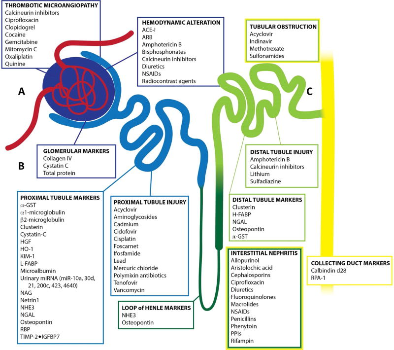
Figure 1. Diagram of renal nephron indicating toxicant, area of injury, and origin of biomarker.
A) Passive filtration is the first process achieved by the glomerulus where toxicant-induced AKI can result in thrombotic microangiopathy and/or hemodynamic alterations. B) In juxtaposition to the glomerulus, the proximal tubule is a primary location of toxicant-induced AKI resulting in proximal tubule injury and loss of integrity leading to downstream accumulation of biomarkers in the urine. C) The remaining tubule structures (Loop of Henle, Distal Tubule, Collecting Duct) are additional structures of the nephron that can become compromised upon toxicant-induced AKI resulting in interstitial nephritis and biomarker accumulation within the urine. (adapted from Casaret and Doull’s Toxicology: The Basic Science of Poisons)

