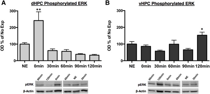
Figure 4.
Phosphorylated ERK activity increases immediately following context exposure in the dHPC while increasing 120 min following context in the vHPC. Rats were given 5 min of context exposure and dHPC and vHPC tissue were collected 0, 30, 60, 90, or 120 min later for Western blotting. Phosphorylation of ERK normalized to β-actin and expressed as a percentage of the No Exposure (NE) control group. (A) Phosphorylated ERK activity significantly increased immediately following context exposure and returned to baseline levels by 30 min. (B) Phosphorylated ERK activity significantly increased 120 min following context exposure. (*) P < 0.05, (**) P < 0.01.

