Figure 4.
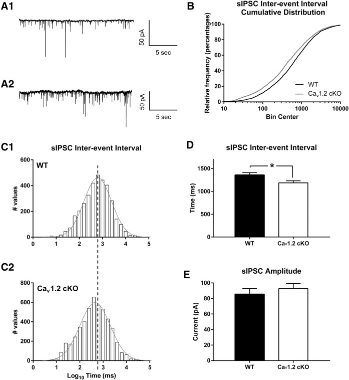
Neuronal deletion of CaV1.2 results in an increase in sIPSC activity in principle neurons of the lateral amygdala. Representative recordings from wild-type (A1) and CaV1.2 conditional knockout mice (A2) of spontaneous IPSCs in inhibition onto principle neurons of the lateral amygdala using whole-cell voltage clamp. (B) CaV1.2 conditional knockout mice exhibited a significant change in sIPSC interevent interval cumulative distribution compared with wild-type mice. (C1,C2) Representation of the sIPSC interevent intervals using a histogram and a fitted Gaussian distribution, showed a leftward shift in CaV1.2 conditional knockout interevent intervals compared with wild-type mice. (D,E) CaV1.2 conditional knockout mice exhibited a significant decrease in the average sIPSC (n = 4884 events in 23 cells from seven mice) interevent interval compared with wild-type littermates (n = 3442 events in 19 cells from six mice), but no change in sIPSC amplitude. Data are represented as mean ± SEM. (*) P < 0.05.

