Figure 1.
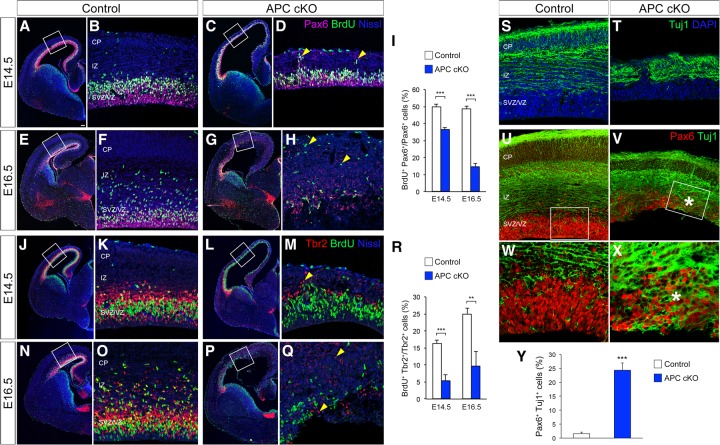
APC deletion disrupts cortical progenitor proliferation and the generation of cortical neurons. (A–H) Proliferating radial progenitors in control (A,B,E,F) and APC cKO (C,D,G,H) cortices at E14.5 (A–D) and E16.5 (E–H) were immunolabeled with anti-Pax6 and anti-BrdU antibodies. (D,H) APC deletion significantly reduced the number of proliferating radial progenitors and led to ectopic collection (arrowheads) of progenitors. (I) Quantification of the number of actively proliferating radial progenitors. (J–Q) Proliferating IPs in control (J,K,N,O) and APC cKO (L,M,P,Q) cortices at E14.5 (J–M) and E16.5 (N–Q) were immunolabeled with anti-Tbr2 and BrdU antibodies. (M,Q) APC deletion resulted in reduced numbers of proliferating IPs and ectopias (arrowheads). (R) Quantification of the number of actively proliferating IPs. A, C, E, G, J, L, N, and P are low-magnification images of cerebral cortices. B, D, F, H, K, M, O, and Q are high-magnification images of dorsal cerebral walls (outlined regions in A,C,E,G,J,L,N,P). (S,T) Generation and placement of Tuj1+ cortical neurons are disrupted in the APC cKO cortex. (U–X) Coimmunolabeling with Pax6 and Tuj1 antibodies indicates an increase in the number of cells that express both Pax6 and Tuj1 (asterisks in V,X) in APC cKO cortex. Images of the cerebral wall (U,V) and high-magnification images of the outlined (box) VZ region (W,X) are shown. (Y) Quantification of the number of Pax6+ Tuj1+ cells. Data shown are mean ± SEM. n = 3 for each genotype. (**) P < 0.01; (***) P < 0.001, Student's t-test. Bars: A,C,E,G,J,L,N,P, 100 µm; U,V, 20 µm; B,D,F,H,K,M,O,Q, 12 µm; S,T, 10 µm; W,X, 5 µm.

