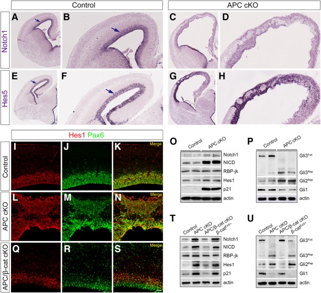
Figure 4.
Notch and Shh signaling pathways are deregulated in the APC-deficient cerebral cortex. (A–H). In situ localization of Notch1 and Hes5 in control and APC cKO cortices indicates disrupted Notch1 and Hes5 expression patterns in APC-deficient cortices (E16.5). (A,B,E,F) Notch1 and Hes5 are highly expressed in the VZ of the cerebral cortex (arrows). Low-magnification (A,C,E,G) and high-magnification (B,D,F,H) images of the cortex are shown. (I–N) Colabeling with Pax6 and Hes1 antibodies shows aberrant expression of Hes1 in APC cKO progenitors. (O) Immunoblot analyses of control and APC cKO cortices reveal up-regulation of Notch1, NICD, Hes1, and p21 levels in APC cKO. (P) An increase in repressor forms of Gli3 and Gli2 and a decrease in Gli1 level are evident in APC cKO. (Q–S) Abnormal Hes1 expression is rescued in the APC/β-catenin double-cKO cortex. (T) Defects in Notch signaling are rescued in the APC/β-catenin double-cKO cortex and recapitulated in the β-catEx3/+ cortex. (U) Abnormal processing of Gli proteins in APC cKO was rescued in the APC/β-catenin double-cKO cortex but not in the β-catEx3/+ cortex. Representative immunoblot images from three biological replicates are shown. Bars: A,C,E,G, 300 µm; B,D,F,H, 100 µm; I–N,Q–S, 40 µm.

