
Figure 11. Combination of two-photon luminescence microscopy and absorbance micro-spectroscopy to characterize nematodes treated with gold nanoparticles.
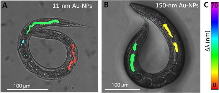
of A) 11 nm Au-NPs and B) 150 nm. The two-photon luminescent signal from Au-NPs is merged with a dark-field micrograph of the treated animals, and colored according to the peak shift of the absorption maxima (by absorbance micro-spectroscopy) compared to the respective Au-NP in dispersion. C) Color legend of the peak shift (expressed in nanometers). Adapted from Gonzalez-Moragas et al.[63]