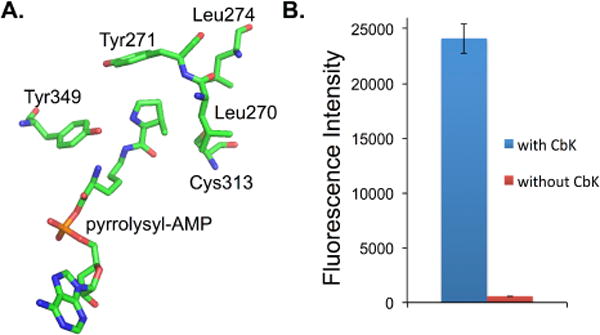
Fig. 2.

Genetic incorporation of CbK. (A) Crystal structure of the PylRS in complex with pyrrolysyl-AMP. The structure is derived from PDB 2ZIM; (B) Fluorescence readings of cells expressing CbKRS and sfGFP-Asn149TAG mutant. The expressions were conducted either in the presence or in the absence of 1.0 mM CbK. Fluorescence intensity was normalized to cell growth.
