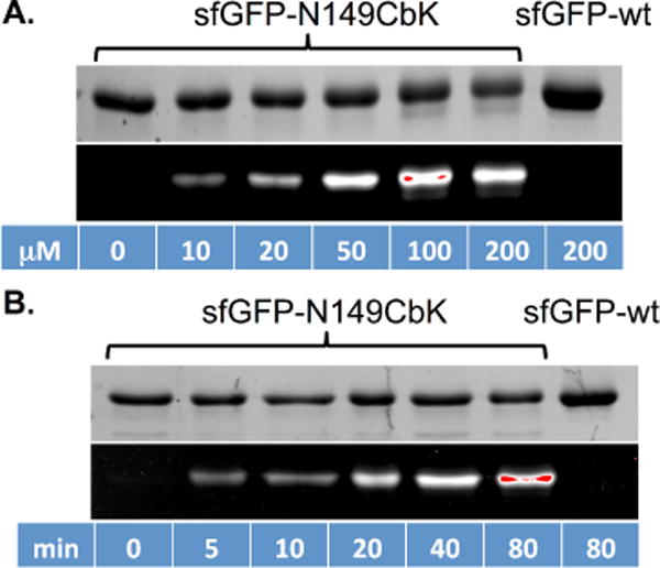
Fig. 3.

In vitro protein labelling of sfGFP variants with cyclobutene-tetrazine reaction. Following labeling reactions, protein samples were denatured by heating, then analyzed by SDS-PAGE. The top panel in each figure shows Coomassie blue stained gel and the bottom panel shows the fluorescent image of the same gel before Coomassie blue treatment. (A) Labeling of sfGFP-N149CbK protein with varied concentrations of Fl-Tet for 80 minutes; (B) Reaction progress of sfGFP-N149CbK protein labeling with 50 μM of Fl-Tet. Wild-type sfGFP was included in both experiments as the control. The fluorescence intensities of these images were quantified and presented in Fig. 7 of ESI. The kobs was estimated as 0.37 M−1 s−1.
