
Figure 8.
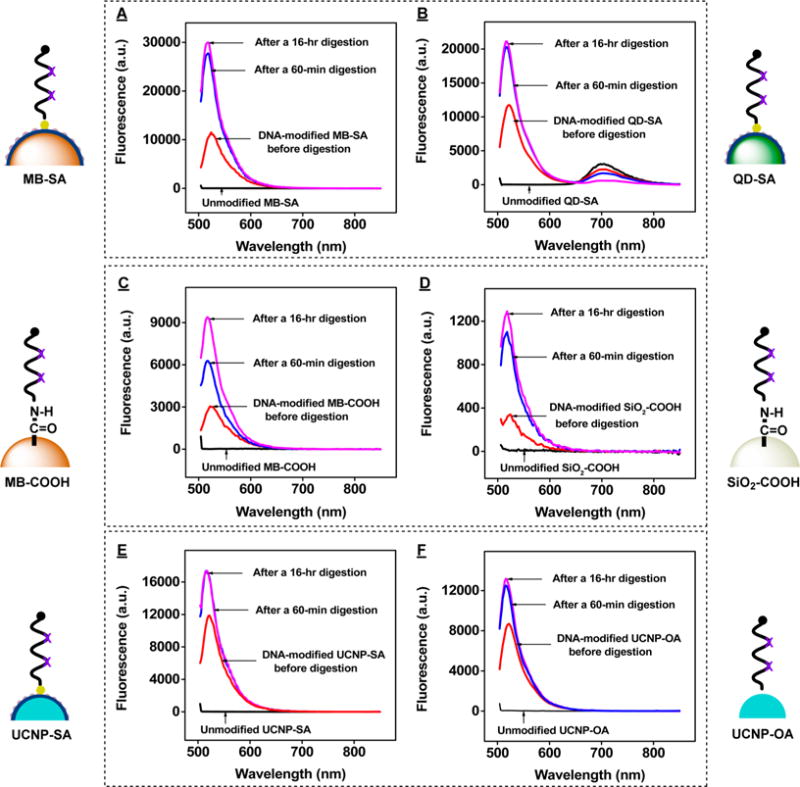
Exo III-based quantification of DNA surface coverage on various particles. Fluorescence spectra of unmodified beads (black), undigested modified beads (red), and supernatants from Exo III-treated, DNA-modified (A) streptavidin-coated magnetic beads (MB-SA), (B) streptavidin-coated quantum dots (QD-SA), (C) carboxylated magnetic beads (MB-COOH), (D) carboxylated silica microspheres (SiO2-COOH), (E) streptavidin-coated UCNPs (UCNP-SA), and (F) oleic acid-capped UCNPs (UCNP-OA). Assays were conducted for 1 h (blue) or 16 h (magenta) with 200 nM cDNA and 0.2 U/μL Exo III.