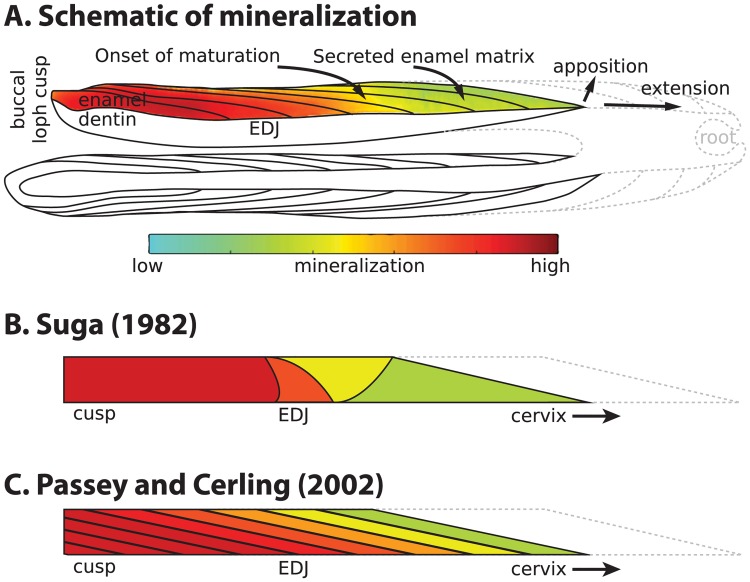
Fig 1. Models of enamel mineralization.
(A) Growing sheep molar viewed in longitudinal section through lingual and buccal (colored) lophs. Mineralization initiates at the dentin horns (left) and proceeds towards the cervix (right) via extension and apposition until crown formation completes. Lines within enamel show incremental addition over time, and colors indicate maturation extent from low (green) to high (red). Solid lines represent formed enamel and dentin, while dotted lines depict future crown outlines. Schematic is based on the first molar (M1) enamel of a 14-week old animal from this study. (B) Mineralization model created by Suga (1982) [15] to represent maturation in large herbivore molars. (C) Mineralization model created by Passey and Cerling (2002) [17] to represent secretion and maturation in ever-growing teeth and tusks.

