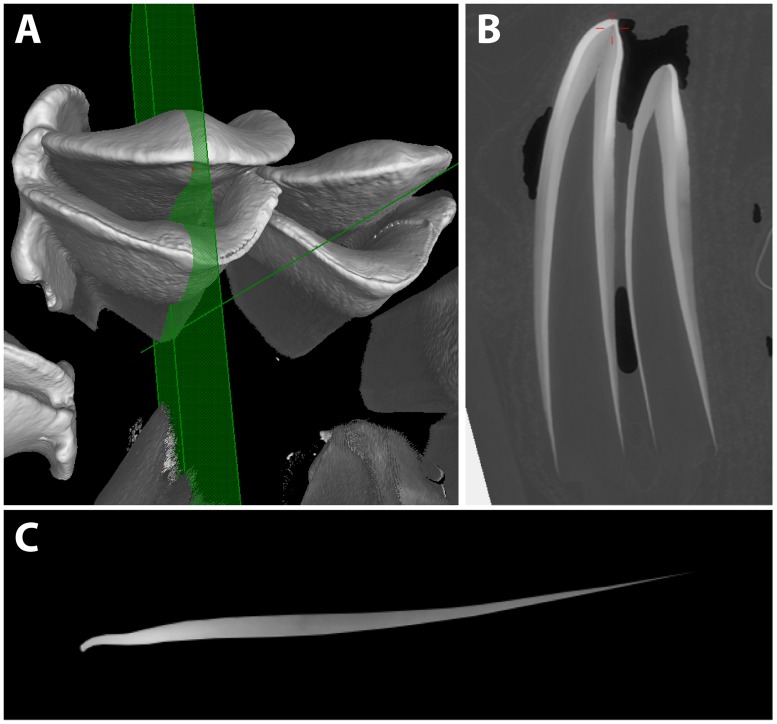
Fig 2. Virtual sectioning of sheep molars for model construction.
(A) Synchrotron-scanned first molar with a bucco-lingual section plane. (B) Resulting section, with the cusp facing upward, lingual loph to the left, and buccal loph to the right. (C) Buccal enamel digitally extracted, with highest mineral density as the brightest shades of grey, and least density in darker shades. Pixel values were measured and converted into hydroxyapatite densities as detailed in the text.

