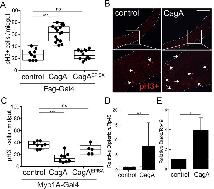
Fig 1. Expression of CagA in adult Drosophila intestinal stem cells (ISCs) promotes proliferation and modulates innate immune components.
(A, B and C) Cell proliferation shown by anti-phospho histone H3+ cells per adult midgut in flies expressing UAS-CagA or UAS-CagAEPISA in (A) intestinal stem cells (ISCs) using esg-Gal4, UAS-GFP or (C) enterocytes (ECs) using Myo1A-Gal4. (B) Representative image from control and ISC expression of CagA (A), anti-phospho histone H3+ shown in red. Scale bar 200 μm. (C and D) qPCR for expression of diptericin (C) and duox (D) in the midgut epithelium of control (esg-Gal4, UAS-GFP) and CagA transgenic Drosophila. *P<0.05, ***P<0.0001 and ns, not significant; for all panels, as assessed by a Student’s t-test. For A and C a representative experiment is shown, each point represents one midgut. n>9 flies/genotype per condition. D and E error bar shows the max expression assayed.

