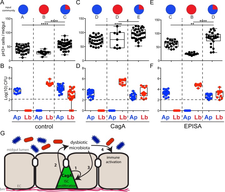
Fig 5. The dysbiotic microbiota of CagA transgenic Drosophila requires interspecies interactions to promote proliferation in the midgut epithelium.
(A-F) Newly eclosed Drosophila reared GF were either mono-associated with Acetobacter pasteurianus (Ap21) or Lactobacillus brevis (Lb22) or community-associated with both Acetobacter pasteurianus (Ap21) shown in red, and Lactobacillus brevis (Lb22) shown in blue, indicated as the input community. (A, C and E) Cell proliferation as shown by anti-phospho histone H3+ cells per midgut. (B, D and F) Microbial abundance of Acetobacter pasteurianus (Ap21) and Lactobacillus brevis (Lb22) observed in the output community seven days post-inoculation. Dashed horizontal line indicates the LOQ. (A and B) Control (esg-Gal4, UAS-GFP), (C and D) CagA (esg-gal4, UAS-GFP; UAS-CagA) and (E and F) CagAEPISA (esg-Gal4, UAS-GFP; UAS-CagAEPISA). *P<0.05 **P<0.01 ***P<0.001 ****P<0.0001, as assessed by a Student’s t-test. Groups were also assessed by ANOVA and shared letters indicate groups that are not statistically different from each other. (G) Model of CagA mediated phenotypes in the adult Drosophila midgut. Expression of CagA in the intestinal stem cells (ISCs) of the adult midgut promotes cell-autonomous proliferation in the midgut epithelium (1), promotes dysbiosis of midgut microbiota (2), and promotes proliferation (3) and overexpression of innate immune factors (4) in a cell non-autonomous manner through dysbiotic microbiota. Enterocytes (EC), basement membrane (bm).

