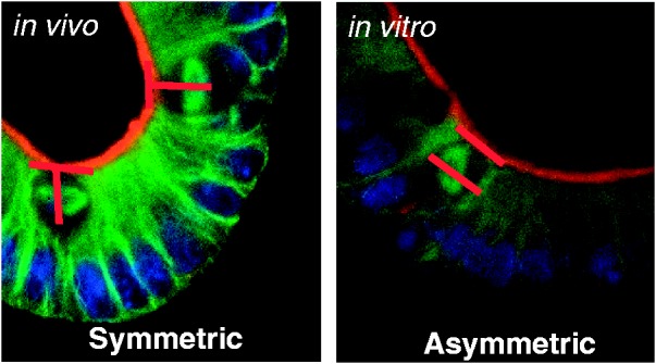
Figure 1.

Representative photomicrograph of symmetric (left) and asymmetric (right) divisions. Green = microtubules (α-tubulin); Blue = nucleus (Dapi); Red = apical border. Note that the chromosome alignment is in an orientation perpendicular to the apical border in symmetric division and parallel in asymmetric. Whereas, the spindle poles are parallel in symmetric division and perpendicular in asymmetric
