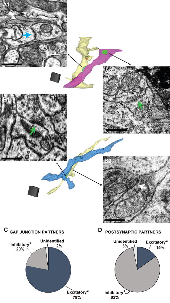
Figure 6. Gap junctional and chemical synaptic partners of putative MC dendrites.
A. A putative MC dendrite (light yellow reconstructed dendrite in middle) connected through a gap junction (A1, teal arrow) to a putative excitatory dendrite (pink reconstructed dendrite), as indicated by the asymmetric dendrodendritic synapse (A2, green arrow) onto another process (not shown in image of reconstructed dendrites). Scale bars = 0.5 µm. Scale cube = 1 µm3. B. Putative MC dendrite (light yellow reconstructed dendrite) that formed a putative excitatory chemical synapse (B1, green arrow) onto a putative inhibitory dendrite (blue reconstructed dendrite), as indicated by the symmetric dendrodendritic synapse (B2, white arrow) onto another process (in this case, it was the same putative MC dendrite). Scale bars = 0.5 µm. Scale cube = 1 µm3. C. Putative MC dendrites formed gap junctions mainly with putative excitatory dendrites. D. Putative MC dendrites formed chemical synaptic contacts mainly onto putative inhibitory dendrites.

