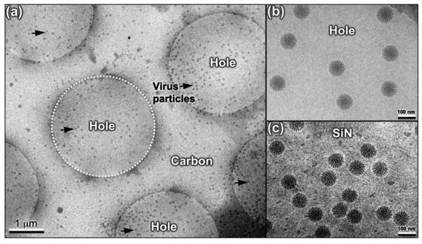
Figure 1. A comparison of rotavirus assemblies prepared using different substrates and methods.

(a) Rotavirus particles (black arrows) are primarily found in holes (white dashed circle) systematically engineered into carbon support films that were plunge-frozen into liquid ethane for cryo-preservation. (b) Close-up view of rotavirus particles located in the holes of carbon support film. (c) Image of rotavirus specimens prepared in the same manner and optimized on silicon nitride (SiN) [7].
