. 2017 Oct 6;6:e26519. doi: 10.7554/eLife.26519

Figure 3. Expression patterns of Trh and 5HT2b genes in the brain.

(A–D) Brains of UAS-mCD8GFP; Trh::Gal4 (A), UAS-stingerGFP; Trh::Gal4 (B), UAS-DscamGFP; Trh::Gal4 (C) and UAS-sytGFP; Trh::Gal4 (D) flies immunostained with the anti-GFP antibody (green) and the neuropil marker nc82 antibody (red). (E–H) Brains of UAS-mCD8GFP; 5HT2b::Gal4 (E), UAS-stingerGFP; 5HT2b::Gal4 (F), UAS-DscamGFP; 5HT2b::Gal4 (G) and UAS-sytGFP; 5HT2b::Gal4 (H), immunostained with anti-GFP antibody (green) and nc82 antibody (red).

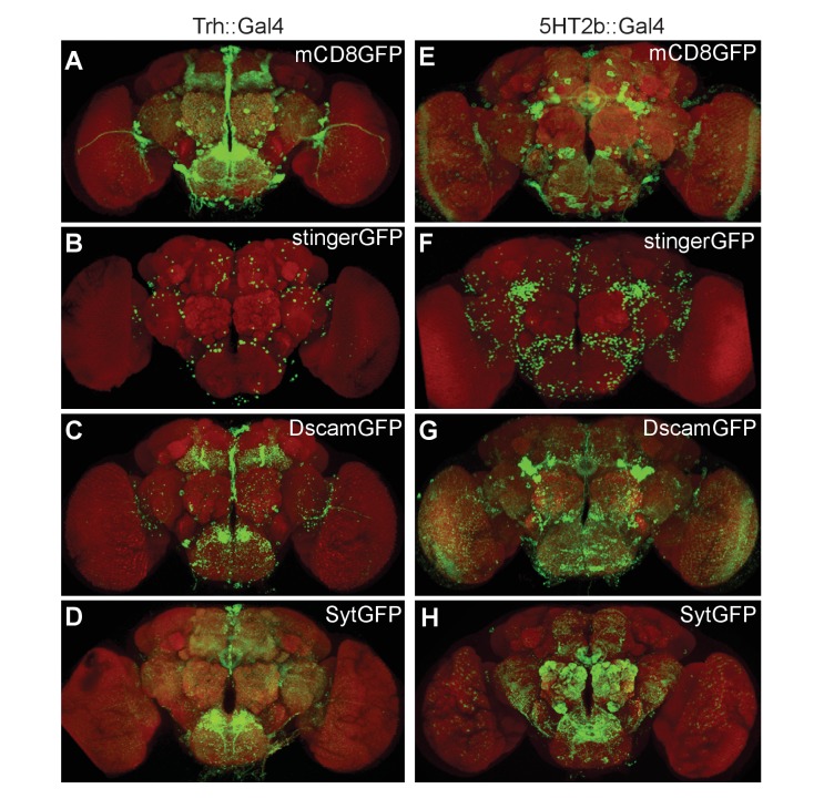
Figure 3.

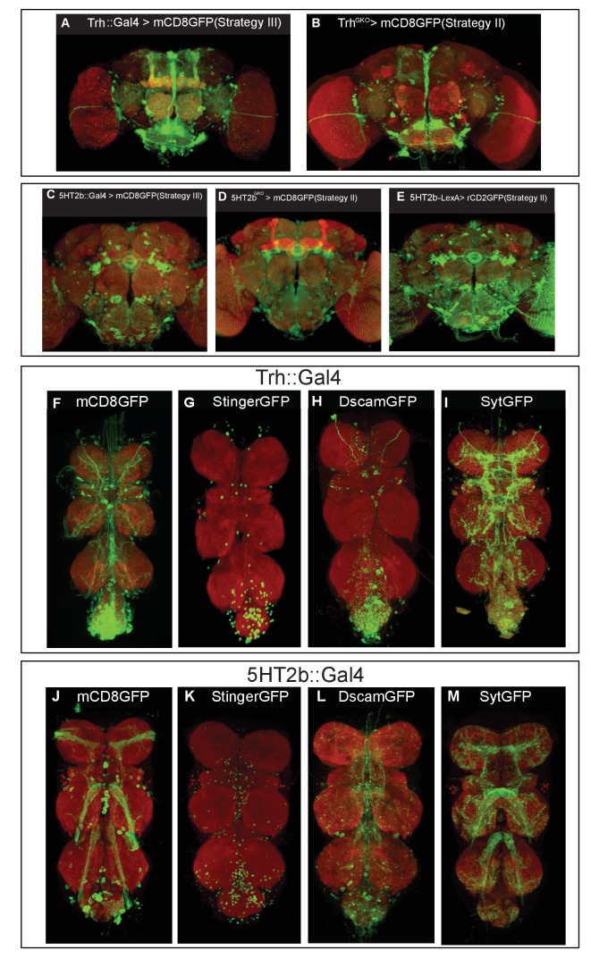
Figure 3—figure supplement 1. Expression patterns of different Trh and 5HT2b strains.

Figure 3—figure supplement 1.

(A–B) Brains of UAS-mCD8GFP; Trh::Gal4 (A) or those of UAS-mCD8GFP; TrhGKO (B) immunostained with the anti-GFP antibody (green) and NC82 (red). Brains of UAS-mCD8GFP; 5HT2b::Gal4 (C), or of UAS-mCD8GFP; 5HT2bGKO (D), or of UAS-rCD2GFP; 5HT2bGKO (E) immunostained with the anti-GFP antibody (green) and NC82 (red). (F–M) Expression patterns of the Trh and 5HT2b genes in the VNC. (F–I) VNCs of UAS-mCD8GFP; Trh::Gal4 (F), UAS-stingerGFP; Trh::Gal4 (G), UAS-DscamGFP; Trh::Gal4 (H) and UAS-sytGFP; Trh::Gal4 (I) flies immunostained with the anti-GFP antibody (green) and NC82 (red). (J–M) VNCs of UAS-mCD8GFP; 5HT2b::Gal4 (J), UAS-stingerGFP; 5HT2b::Gal4 (K), UAS-DscamGFP; 5HT2b::Gal4 (L) and UAS-sytGFP; 5HT2b::Gal4(M) flies immunostained with the anti-GFP antibody (green) and NC82 (red).