
Figure 1. Murine allograft model of ovarian cancer metastasis demonstrates peritoneal seeding by cancer cells/MCAs with subsequent penetration and remodeling of sub-mesothelial collagen.
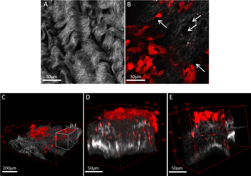
C57Bl/6 female mice were injected intraperitoneally with ID8-RFP murine EOC cells and sacrificed at 8–10 weeks post injection. The parietal peritoneum was dissected and prepared for combined fluorescence/SHG microscopy as described in Methods. Shown are examples of (A) tumor-free mouse peritoneal explant (collagen, grey) and (B) peritoneal explant (collagen, grey) containing a metastatic lesion (cancer cells, red) exhibiting collagen reorganization and peri-cellular collagen clearance areas (arrows). Scale bars: as indicated. Murine metastatic lesions depict (C) seeding of cancer cells and cell clusters (red) atop of peritoneal collagen layer (grey), (D) 3D volume view and (E) orthoslice view of cancer cells penetrating the sub-mesothelial collagen layer. Scale bars: as indicated.