
Figure 7. Inhibition of MT1-MMP modulates matrix invasion and collagen remodeling by EOC cells.
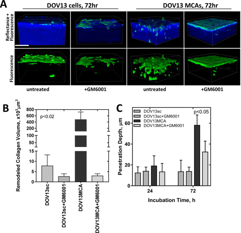
A) Individual Ncad+ DOV13 cells (that endogenously express MT1-MMP) were applied atop 3D collagen gels (1.5mg/ml) inside a glass-bottom dish, and incubated with the broad spectrum MMP inhibitor GM6001 (25μM) or with no inhibitor, as detailed in Methods. Nikon A1R-MP confocal microscope was used for continuous z-stack imaging of cancer cells (green, fluorescence mode) and collagen (blue, reflectance mode). Representative images at 72h incubation time point are shown. Scale bar: 100μm. Evaluation of (B) peri-cellular collagen remodeling after 72hr of incubation, and (C) cell penetration depth at 24 and 72h. All assays were repeated in triplicate and statistical analysis was performed using Mann-Whitney U test. Statistical significance is shown for Ovca433MT1-MMP and OvCa433Ncad+ with respect to OvCa433 invasiveness.