
Figure 4. EMP2 promotes tumor growth in Ishikawa cells.
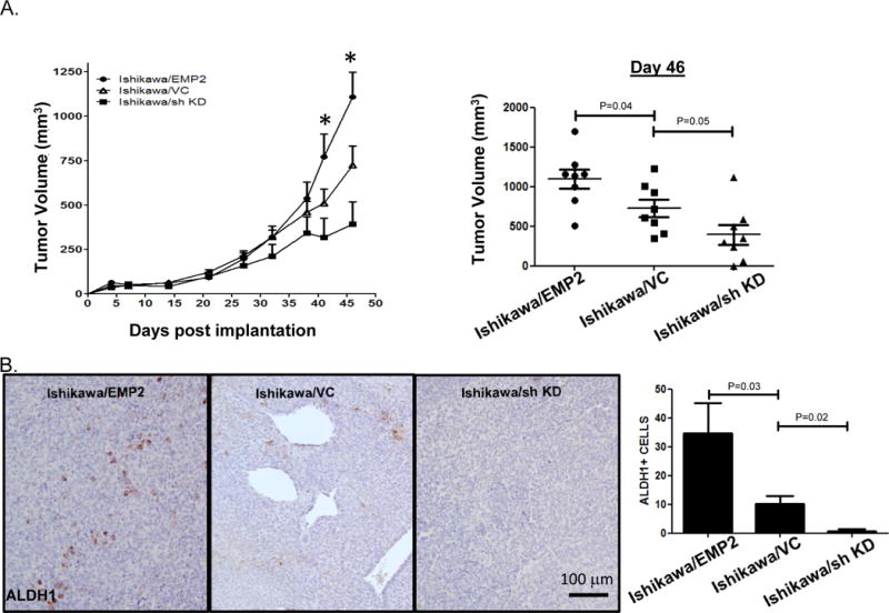
A. 1×10⁶ Ishikawa cells with modified EMP2 levels (Ishikawa/EMP2, Ishikawa/VC, and Ishikawa/sh KD) were injected into injected s.c. into BALB/c Fox Chase SCID mice, and tumor growth monitored for 46 days. Two way ANOVA, p<0.05. Tumor volume on day 46 is shown to the right with groups compared using Student’s t test. B. Tumors were then harvested and ALDH1 expression measured using IHC. Magnification, 200X. ALDH+ cells were enumerated in 4 independent fields from at least 3 tumors with results depicted as the average ± SEM shown to the far right.