
Figure 7. Anti-EMP2 IgG1 reduces ALDH expression in vitro.
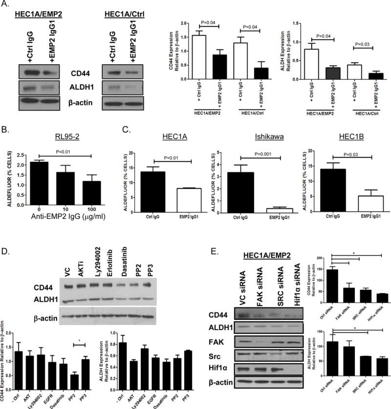
A. HEC1A/VC or HEC1A/EMP2 cells were treated with anti-EMP2 IgG1 or human IgG at 50μg/ml for 24 hrs. Left, representative western blots showing ALDH1 and CD44 expression relative to β-Actin. Right, relative expression of ALDH1 and CD44 expression from three independent experiments normalized to β-Actin. B. RL95-2 cells were treated with 10 or 100μg/ml of anti-EMP2 IgG1 for 24 hours. ALDH activity was measured using the ALDEFLUOR assay. C. HEC1A (left), Ishikawa cells (middle), and HEC1B (right) were treated with control or anti-EMP2 IgG1 antibody with 50μg/ml. ALDEFLUOR activity was measured after 24 hours with the average activity from three independent experiments ± SEM. D. HEC1A/EMP2 cells were treated with common inhibitors targeting the AKT, PI3K, EGFR, and FAK/Src, pathways under hypoxic conditions for 24 hours. PP3 as well as a vehicle control (saline) were included as controls. Total CD44 and ALDH1 levels were measured using western blot analysis and a representative graph is included. Right, Levels of CD44 and ALDH1 expression were quantitated to relative to β-Actin from three independent experiments. Results are presented as the mean ±SEM. *, p<0.05. E. HEC1A/EMP2 cells were treated with FAK, Src, and HIF-1α siRNA under hypoxic conditions. CD44, ALDH1, FAK, Src, HIF-1α, and β-Actin levels were measured using western blot analysis with a representative graph included on the left. Right, Levels of CD44 and ALDH1 expression were quantitated to relative to β-Actin from three independent experiments. Results are presented as the mean ±SEM.