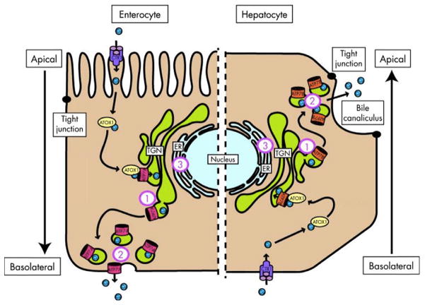
Fig. 3.1.
Schematic representation of copper-induced relocalization of ATP7A and ATP7B. The left side of the diagram represents an enterocyte and the right side represents a hepatocyte. On both sides, copper enters the cell through copper transporter 1 (CTR1) and is escorted by copper chaperone antioxidant protein 1 (ATOX1) to ATP7A or ATP7B in the trans-Golgi network (TGN). When copper levels rise above a certain threshold, ATP7A and ATP7B excrete copper into the plasma on the basolateral side of the enterocyte and into the bile on the apical side of the hepatocyte. Defects in localization of ATP7B may lead to copper accumulation at the (1) TGN due to unresponsiveness, (2) cell periphery, and (3) endoplasmic reticulum (ER) due to misfolding. (Reproduced from de Bie et al., 2007.)

