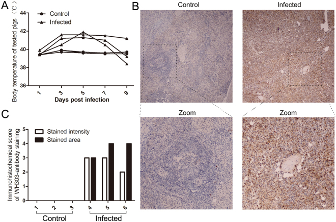
Figure 1.
Body temperature of tested pigs and distribution of the CSFV antigen in the spleen. (A) Body temperature of tested pigs. 3 pigs were infected, and 3 pigs were used as controls. Each infected pig was intramuscularly injected with 105 TCID50 of Shimen strain CSFV. Body temperature was assessed every 2 days until 9 days post infection. (B) Location of CSFV in the spleen. Paraffin sections of spleens were stained with mouse monoclonal anti-CSFV E2 protein antibody (1:50) as described in Materials and Methods. The images were a representative immunocytochemistry result captured at magnifications under 20× objectives. (C) Immunocytochemistry score. 3 sections of each sample were prepared, and 3 random visual fields of each section were snapped. The score was evaluated by two different clinical doctors. Score of stained intensity: characteristics of positive-stained cells. Pale yellow, tan and sepia were scored as 1, 2, and 3, respectively. Score of stained area: the mean ratios of positive cells in each visual field. Scores of 1, 2, 3, and 4 were corresponded to 6~25%, 26~50%, 51~75% and >75%, respectively.

