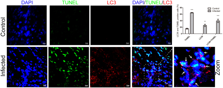
Figure 4.
Confocal microscopy showing the connection of autophagic and apoptotic cells. Frozen sections stained with TUNEL (green) were simultaneously immunostained with LC3II-antibody (red). Nuclei was stained with DAPI. In the zoomed images, the arrows indicated the colocalization of cells both undergoing autophagy and apoptosis. The average ratios of TUNEL-positive, LC3II-positive and double stained cells were based on at least 50 cells in each group (mean ± SD; n ≧ 50 cells; **p < 0.01, ***p < 0.001).

