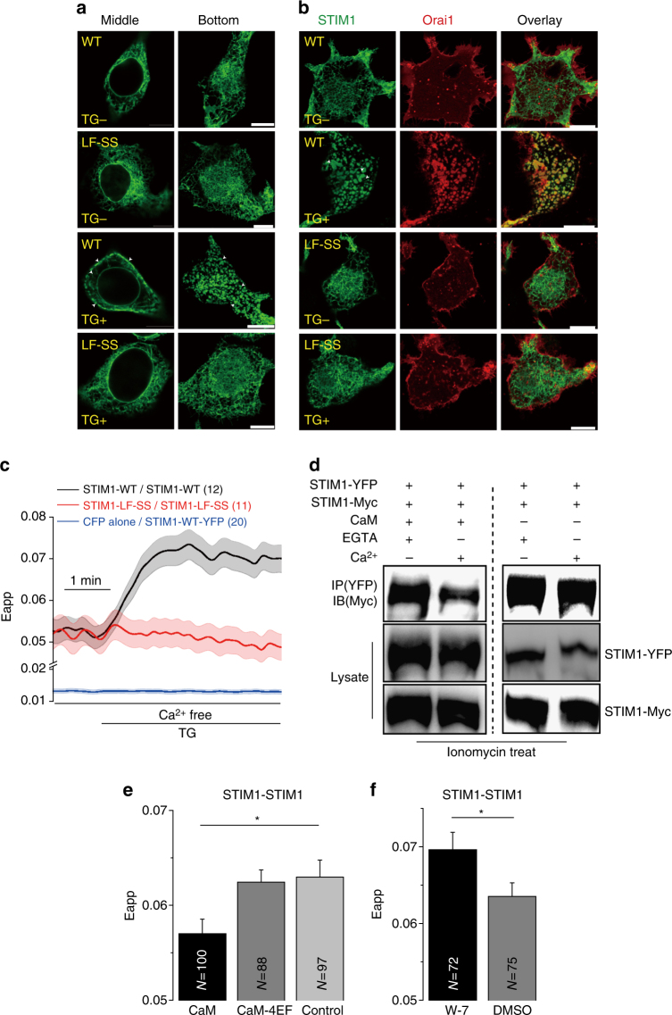
Fig. 6.
Ca2+-CaM disassembles the STIM1 oligomer. a, b Confocal images of live HEK293T cells that were transfected with STIM1-GFP (WT or mutant) alone or co-transfected with Cherry-Orai1 before and after 1 μM TG stimulation. Puncta are indicated by the arrow. Scale bars are 10 μm. c FRET was measured between WT or mutant with COOH-terminus CFP- and YFP-tagged STIM1 in HEK293T cells. TG (1 μM) was used to induce Ca2+ depletion. d Western blot analysis of Ca2+-CaM protein competitive binding with STIM1 intermolecular interactions in HEK293T cells. Ionomycin (3 μM) was used to induce Ca2+ depletion before lysis. e Steady-state FRET between STIM1-CFP and STIM1-YFP that were co-expressed with CaM or CaM-4EF mutant in HEK293T cells after 3 μM ionomycin treatment. f Steady-state FRET between STIM1-CFP and STIM1-YFP pretreated with 30 μM W-7 or DMSO control for 30 min after 3 μM ionomycin treatment in HEK293T cells. Numbers of cells analyzed are indicated. *P < 0.05 (unpaired Student’s t-test). Error bars denote SEM

