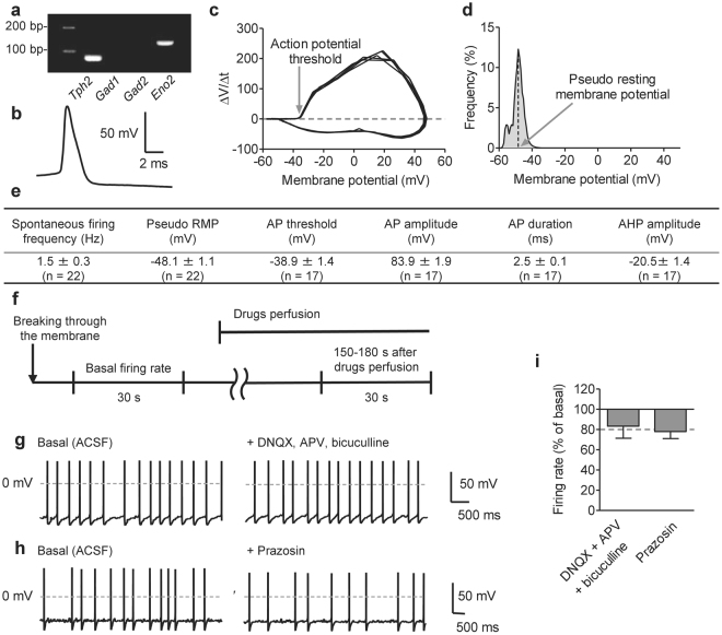
Figure 1.
Serotonergic neurons in the dorsal raphe nucleus (DRN) show spontaneous firing activity. (a) Representative cropped image of single-cell reverse transcription polymerase chain reaction after whole-cell recording. Tryptophan hydroxylase 2 (Tph2) mRNA was used as a marker of serotonergic neurons. Glutamate decarboxylase 1 and 2 mRNA (Gad1 and Gad2), markers of GABAergic neurons, were used as negative controls. Gamma-enolase mRNA (Eno2), a marker for neurons, was used as a positive control. Uncropped image was shown in Supplementary Fig. S1. (b) Representative trace of the action potential (AP) recorded from DRN serotonergic neurons. Serotonergic neurons showed a wide action potential and a long-lasting after hyperpolarization. (c) Representative phase plane plot of membrane potential vs. its derivative with respect to time (dV/dt). Five APs from one neuron was plotted. (d) Representative membrane voltage histogram. The higher voltage peak was considered as pseudo resting membrane potential (RMP). (e) Electrophysiological characters of 22 serotonergic neurons from 7 mice. Recordings were performed in normal ACSF condition without any drug or electrical stimulation. AHP; afterhyperpolarization. (f) Time course of recording the effects of drug perfusion. Spontaneous firing was recorded for 30 s before and after drug application, and changes in the firing rate were calculated. (g,h) Representative traces of the spontaneous firing before (left) and after (right) the application of DNQX (20 μM), APV (50 μM) and bicuculline (20 μM) (g) or prazosin (1 μM) (h). (i) The changes in the spontaneous firing rate before and after the application of DNQX (20 μM), APV (50 μM), and bicuculline (20 μM), or prazosin (1 μM). (DNQX + APV + bicuculline, n = 4 neurons from 3 mice, P = 0.2545 by paired t-test; prazosin, n = 3 neurons from 2 mice, P = 0.0855 by paired t-test). Data are presented as the mean ± S.E.M.

