Fig. 2.
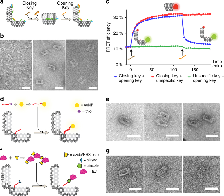
Reversible closing–opening mechanism and cargo loading. a Schematic of the closing–opening mechanism of the DNA nanostructure. b Transmission electron microscopy (TEM) images showing open, closed and reopened DV samples. Original TEM images in Supplementary Fig. 6. c FRET efficiency measured on the open Cy3/Cy5-conjugated DV. The blue line shows values measured after the closing key is added (brown), and—subsequently—after the opening key is added (orange). d Schematics of non-covalent cargo-loading mechanism: 5 nm gold nanoparticles (AuNPs) were reacted with thiol-modified DNA strands and incubated together with open DV molecules exposing a complementary CAS staple strand within the cavity. e TEM images of AuNP-loaded DV. AuNPs are detectable as black dots within the DV cavity. Original TEM images in Supplementary Fig. 10a–c. f Schematics of covalent cargo-loading mechanism: the endopeptidase aCt was conjugated with azide-NHS ester handles, and then reacted together with open DV exposing an alkyne-modified CAS staple strand within the cavity. Copper(I)-catalysed alkyne-azide cycloaddition reaction induces the formation of covalent DNA origami-enzyme conjugates. g TEM images of the aCt-loaded DV after incubation with closing key. Enzymes are detectable as white spots within the DV cavity. Original TEM images in Supplementary Fig. 10d–f. Scale bar, 50 nm

