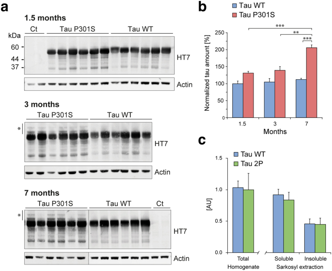
Figure 3.
The level of human tau increases over time in the brain of mice injected with AAV-P301S. (a) Detection of overexpressed tau using the human tau-specific HT7 antibody in mouse forebrain homogenates at 1.5, 3 and 7 months post-AAV injection. Actin is used as loading control. *Indicates higher molecular weight human tau species observed in the P301S group of mice. (b) Relative human tau levels measured by western blotting of mouse forebrain homogenates (1.5, 3 and 7 months post- injection). Blots are shown in Supplementary Figure 1. For each animal, human tau values are normalized to actin and plotted as percentage of the mean value for the AAV-WT group at 1.5 months. Statistical analysis: two-way ANOVA followed by Tukey’s post-hoc test, group × time effect F(2,30) = 4.89, **p < 0.01; ***p < 0.001. (c) The graph compares the level of human tau (HT7/TAU-13 AlphaLisa) in total soluble proteins from forebrain homogenates, as well as in the soluble and insoluble fractions following sarkosyl extraction. Samples are derived from AAV-WT and AAV-2P mice, 7 months after AAV injection; n = 8 mice per group.

