Figure 3.
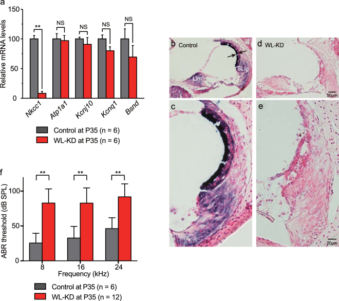
Quantitative, qualitative, and functional analyses of lifetime Nkcc1 knockdown effects in Actin-tTS::Nkcc1 tetO/tetO mice (WL-KD mice). (a) At P35, cochlear Nkcc1 transcription is decreased in WL-KD mice vs. controls in comparison with other K+ circulation molecules as labeled. mRNA levels are normalized to Gapdh. Nkcc1 mRNA was significantly suppressed in WL-KD mice compared with controls, but the mRNAs of other K+ circulation molecules were not suppressed. (b–e) In situ hybridization of Nkcc1 mRNA in mouse cochleae. (b) Normal Nkcc1 mRNA expression in cochlea of control. Arrows show normal SV thickness. (c) Magnification of view in (b). (d) Strong suppression of Nkcc1 level in WL-KD mice. (e) Magnification of view in (d). (f) Nkcc1 knockdown significantly elevates the ABR threshold, which indicates hearing loss. **Significant difference at p < 0.01 (Mann-Whitney test). NS: no significant difference (Mann-Whitney test).

