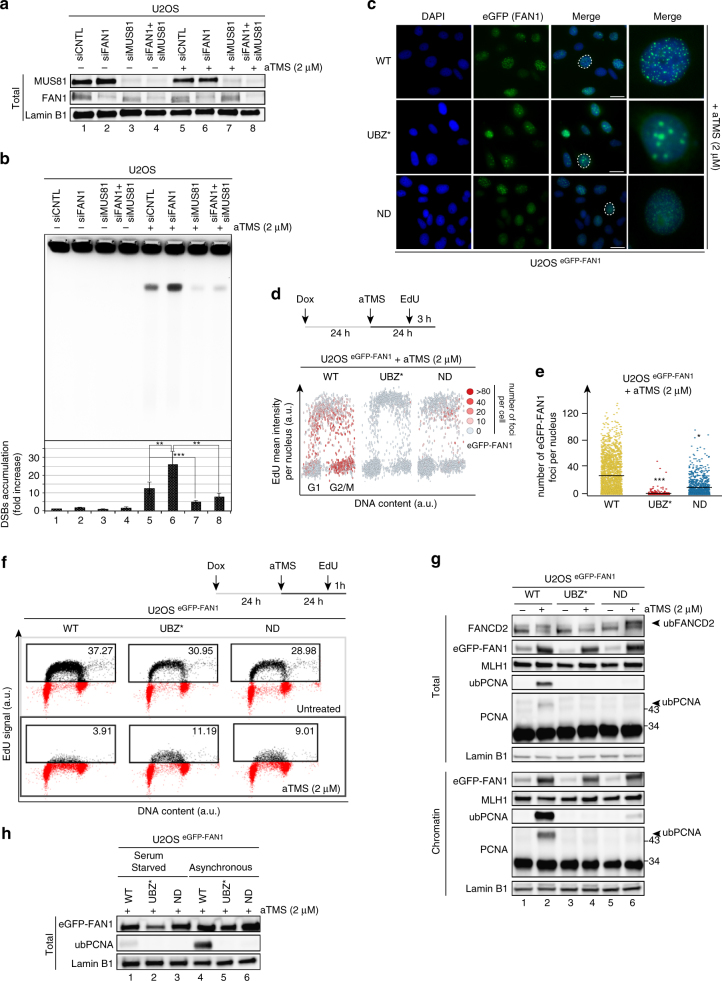
Fig. 1.
FAN1 prevents fork collapse and limits DNA synthesis by promoting PCNA ubiquitylation. a Immunoblot of extracts of U2OS cells transfected with siRNAs against FAN1 and/or MUS81, and treated or mock-treated with aTMS (2 μM; 48 h). The antibodies used are shown on the left. The figure shows a representative blot of three independent experiments. b PFGE of DNA isolated from cells treated as described in a was performed to visualize DSB induction. A quantification of three independent experiments is shown. Data are represented as mean ± s.d. (n = 3). Statistical analysis was carried out using unpaired, two-tailed t-tests. P values expressed as **(P < 0.01) or ***(P < 0.001) were considered significant. c Representative images of U2OS cells expressing the indicated eGFP-FAN1 variants after treatment with aTMS (2 μΜ; 24 h). Scale bar: 25 μm. d Quantitative image-based cytometry (QIBC) of eGFP-FAN1 foci in cells from c pulse-labeled with EdU during the last 3 h of aTMS treatment. The heat map indicates the mean eGFP-FAN1 intensity per nucleus. e Quantification of eGFP-FAN1 foci count derived from the QIBC analysis in d. Median levels are indicated by black bars. Statistical analysis was carried out using unpaired, two-tailed t-tests. P values expressed as ***(P < 0.001) and *(P < 0.05) were considered significant, n = 3. f Cells as in c were treated with EdU (1 h) and Click chemistry. EdU incorporation was evaluated by FACS. g Total cell extracts and chromatin-enriched fractions of cells as in c were analysed by immunoblotting using the antibodies shown on the left. A representative blot of four independent experiments is shown. h Immunoblot analysis of total cell extracts of exponentially-growing or serum-starved U2OS cells expressing the indicated eGFP-FAN1 variants following treatment as in c. A representative blot of two independent experiments is shown

