Fig. 6.
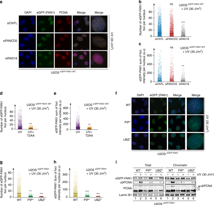
The FAN1 PIP-box motif is required for FAN1 foci formation upon UV exposure. a U2OS cells expressing eGFP-tagged FAN1 WT were transfected with the indicated siRNAs and treated or mock-treated with UV (30 J/m2, 4 h release) before immunostaining with anti-PCNA antibody. Representative images are shown. Scale bar: 25 μm. b, c Quantification of eGFP-FAN1 foci count (b) and the sum of their intensities (c) derived from QIBC analysis of a. Median levels are indicated by black bars. Statistical analyses were carried out using unpaired, two-tailed t-tests. P values expressed as ***(P < 0.001) and **(P < 0.01, n = 3) (d, e) Cells as in a were treated with UV (30 J/m2, 4 h release) and incubated or not with T2AA (40 μΜ; 6 h). Quantification of eGFP-FAN1 foci count (d) and the sum of their intensities (e) was obtained from QIBC analysis. Median levels are indicated by black bars. Statistical analysis was carried out using unpaired, two-tailed t-tests. P values expressed as **(P < 0.01) were considered significant (n = 3). f Representative images of U2OS cells expressing the indicated eGFP-FAN1 variants after treatment with UV (30 J/m2, 4 h release). Scale bar: 25 μm. g, h Quantification of eGFP-FAN1 foci count (g) and the sum of their intensities (h) was obtained from the QIBC analysis of f. Median levels are indicated by black bars. Statistical analyses were carried out using unpaired, two-tailed t-tests. P values expressed as ***(P < 0.01) or **(P < 0.01) were considered significant, n = 3. i Total cell extracts and chromatin-enriched fractions of U2OS cells expressing the indicated eGFP-FAN1 variants and treated or mock-treated with UV (30 J/m2, 4 h release) were analysed by immunoblotting using the indicated antibodies. A representative blot of four independent experiments is shown

