
Figure 1.
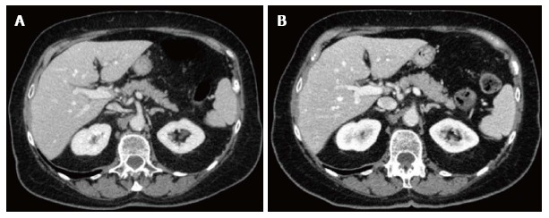
Axial contrast-enhanced computed tomography images at the level of upper abdomen obtained in a 81 years old female patient with lung cancer (height 160 cm, weight 61 kg). A: Standard dose protocol (120 kV, 300 mAs, DLP 1317.4 mGy*cm, CDTI 21.1 mGy); B: Lower dose protocol (120 kV, 142-222 mAs, DLP 846.0 mGy*cm, CDTI 13.6 mGy): Lower dose image shows increased sharpness and enhancement in comparison with standard dose image in spite of mild increase of noise, and similar diagnostic quality with a 35.8% Dose-Length-Product reduction.